BE AWARE: We only accept payment at the hospital reception. Don't respond to unknown emails, phone calls, or any app downloads for any online payment. Do not share OTP; we don't encourage payment via calls and apps.

Patient Education Material

PATIENT EDUCATION

Information in this segment has been assembled to help you better understand the anatomy and function of the knee and the degenerative changes associated with arthritis, to prepare you for total knee replacement surgery, and to provide guidelines for post operative care. The long term goal of total knee replacement surgery is to provide relief of pain, restore normal activities of daily living and enhance your quality of life.

Anatomy and Function of the Knee

The three bones that comprise the knee joint are the femur (thighbone), the tibia (shinbone), and the patella (kneecap). The knee may be described as a modified hinge joint, similar to the hinge on a door. However, the knee not only bends back and forth like a hinge, it has a complex rotational component that occurs with flexion and extension of the knee. The knee is a major weight-bearing joint that is held together by muscles, ligaments and other important soft tissue. Cartilage is the material inside the joint that provides shock absorption the knee during weight-bearing activities such as walking or stair climbing.

Anatomy and Function of the Knee
Arthritic Knee

The Arthritic Knee

Arthritis in the knee joint occurs as a result of degeneration of the cartilage in your knee. Osteoarthritis is commonly referred to as “wear and tear” arthritis and is the most common cause for total knee replacement surgery. Due to osteoarthritis, the cartilage in the knee breaks down over time and the result is a severely damaged joint surface with bone rubbing on bone. This process may occur as a result of previous trauma to the joint, ligament instability, or abnormal stresses to the joint. Rheumatoid arthritis is an inflammatory process that results in erosion of the articular cartilage and subsequent damage to the knee joint surface.

Non-Surgical Treatment of the Arthritic Knee

Listed below are several non-operative, conservative options to consider for treatment of the arthritic knee.

  • Lifestyle Modification: Losing weight, avoiding aggravating activities, modifying exercise to low impact activities only.
  • Exercises: Specifically prescribed exercises to improve strength and flexibility without exacerbating your pain.
  • Anti-inflammatory Medications: Designed to decrease swelling in the joint and provide temporary pain relief.
  • Corticosteroid Injection: Powerful anti-inflammatory agent injected directly into the joint.
  • Joint Fluid Therapy: A series of injections directly into your knee, designed to improve lubrication in the joint.
  • Glucosamine/Chondroitin: Dietary Supplement that may relieve arthritic pain.
  • Bracing: Used to provide external stability to the knee joint.
  • Arthroscopic Surgery: Minimally invasive procedure to remove debris or repair torn cartilage.

Reasons For Knee Replacement Surgery

Total knee replacement surgery is considered when all other conservative meas¬ures have failed to provide successful intervention and may be performed for the following reasons:

  • To relieve pain
  • To improve joint stability
  • To improve alignment and correct bone deformity
  • To maximize quality of life
  • To optimize activities of daily living

Total knee replacement surgery is a common procedure performed on more than 600,000 people worldwide each year. With recent advancements in surgical tech¬nique and implant design, patients have experienced dramatic improvement in knee pain, function, and quality of life. Furthermore, most patients can now expect their implants to last up to a decade or more, allowing for years of active, healthier, pain-free living.

Preparation For Knee Replacement Surgery

Once you and your orthopaedic surgeon have decided to proceed with surgery, there are several activities that must occur prior to surgery, including the following:

  • Initial Surgical Consultation: Preoperative X-rays, complete past medical history, complete past surgical history, complete list of all medications and allergies (prescription, over-the-counter, vitamin supplements).
  • Complete Physical Examination: Your internist or family physician will determine if you are in the best possible condition to undergo surgery.
  • Blood Donation: A transfusion may be necessary after surgery, therefore you may wish to donate blood prior to surgery.
  • Physical Therapy: Instruction in an exercise program to begin prior to surgery and an overview of the rehabilitation process after surgery will better prepare you for post-operative care.

Preparation for the Hospital: you may want to bring the following items:

  • Clothing: underwear, socks, t-shirts, exercise shorts for rehabilitation
  • Footwear: walking or tennis shoes for rehab; slippers for hospital room
  • Walking Aids: walker, cane, wheelchair or crutches if used prior to surgery Insurance Information

Evening Before Surgery:

  • Do not eat or drink after midnight.
  • Shower (with 5-minute scrub to surgical area with special soap provided).
  • Prepare your belongings and review total knee replacement information.

Preoperative Procedures

This section will give you a brief overview of the activities that will occur on the day of surgery:

  • You will be admitted to the hospital, typically the morning of your surgery.
  • A final assessment of vital signs will be taken.
  • A clean hospital gown will be provided.
  • An IV will be started to give you fluids and medication during and after the procedure.
  • An elastic stocking may be provided to decrease the likelihood of blood clots.
  • You will be asked to empty your bladder.
  • All jewelry, dentures, contacts, and nail polish must be removed.
  • The surgical leg will be scrubbed and shaved in preparation for surgery.
  • The anesthesiologist will discuss the type of anesthesia that will be used.
  • You will be taken into the operating room.

Total Knee Replacement

 
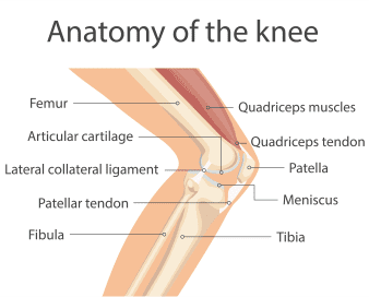
In the knee replacement procedure, each prosthesis is made up of four parts. The tibial component has two elements and replaces and the top of the shin bone or tibia. This prosthesis is made up of a metal tray attached directly to the bone and a high-density plastic spacer that provides the bearing surface. The femoral component replaces the bottom of the thigh bone or femur. This com¬ponent also replaces the groove where the patella or kneecap rides. The patellar component replaces the surface of the knee cap, which rubs against the femur. The patella protects the joint, and the resurfaced patellar button will slide smoothly on the front of the joint. In some cases, surgeons do not resurface the patella.

One of the keys to a successful implant is its ability to withstand the rigors of daily activity, and central to that is the quality of the artificial surfaces that slide against each other, or articulate, in the new joint. In knee implants, bearing surface options have been somewhat limited over the last few decades. The standard substance used for the femoral component is cobalt chrome, a metal alloy typified by its tough¬ness and biocompatibility. However, even this high-quality industry standard has its shortcomings. Over time, this metal surface can become roughened by bone and bone cement particles trapped between the femoral component and the plastic tibial insert.

This roughened surface, when rubbing against the plastic component up to two million times per year, can more quickly wear out your implant. When that happens, you will have to undergo surgery to replace the plastic piece, the femoral compo¬nent, and possibly even the tibial component. For this reason, implants have been shown to last between 10 and 15 years in the human body. The most exciting material to enter orthopaedics in recent years is OXINIUM° (oxi¬dized zirconium.)This remarkable new material combines the strengths of ceramic and metal, such as wear-reduction and strength, but does not have the weakness¬es, such as limited implant options and the possibility of fracture.

Zirconium is a biocompatible metal, similar to titanium. When the zirconium alloy undergoes a unique heating process, the surface of the metal transforms into a ceramic. Even though the new ceramic surface is 4,900 times more abrasion resistant than cobalt chrome, it retains the toughness and flexibility of the underlying metal. Because it can achieve this remarkable reduction in implant wear without sacri¬ficing strength as actual ceramic com¬ponents do, OXINIUM° implants have the potential to last significantly longer, thus possibly reducing the need for future corrective surgeries. When articulating on standard plastic tibial components, OXINIUM° knee implants reduce wear by 85-percent compared to cobalt chrome.

Knee replacement surgery typically takes between one and two hours to complete. This section will provide you with a brief, easy-to-understand description of the surgical procedure:

  • An incision is made extending from the thigh, past the inside edge of the kneecap, and down to the shinbone.
  • The end of the femur is shaped in preparation for sizing the femoral trial component.
  • The top of the tibia is shaped for proper sizing of the tibial trial component.
  • The trial units are put in place and the appropriate implant size is selected.
  • The knee is assessed for align¬ment, stability, and range of motion.
  • The underside of the kneecap is prepared and patella trial is selected.
  • The trial units are removed and the final femoral, tibial, and patella components are implanted.
  • The incision is closed, a drain is put in and the post-operative bandaging is applied.

After your surgery is completed, you will be transported to the recovery room for close observation of your vital signs, circulation, and sensation in your legs and feet. As soon as you awaken and your condition is stabi¬lized, you will be transferred to your room. Below is an example of what you may see when you wake up:

  • You will find a large dressing applied to your incision in order to maintain cleanliness and absorb any fluid.
  • There may be a drain placed near your incision in order to record the amount of drainage being lost from the wound.
  • You may be wearing elastic hose and/or a compression stocking sleeve designed to minimize the risks of blood clots.
  • Your doctor may prescribe a PCA (patient-controlled analgesia) that is connect¬ed to your IV. The unit is set to deliver a small, controlled flow of pain medica¬tion and is enacted when you firmly press the button on your machine. Press the button anytime you are having pain.
  • You may have a catheter inserted into your bladder as the side effects of anes¬thesia often make it difficult to urinate.

Preventing Complications

In a small percentage of patients, as with all major surgical procedures, complica¬tions can occur. Below is a list of potential complications and steps you can take to prevent their occurrence:

Thrombophlebitis

Thrombophlebitis: also known as deep vein thrombosis (DVT), this problem occurs when the large veins of the leg form blood clots and in some instances, become lodged in the capillaries of the lung and cause a pulmonary embolism. The following steps may be taken to avoid blood clots:

  • Blood-thinning medication (anticoagulants)
  • Elastic stockings (TED hose)
  • Foot and ankle exercises to increase blood flow and enhance venous return in the lower leg.

Important: If you develop swelling, redness, pain, and/or tenderness in the calf muscle, report these symptoms to your orthopaedic surgeon or internist immediately.

Infection

Although great precaution is taken before, during, and after surgery, infections do occur in a small percentage of patients following knee replacement surgery. Steps you can take to minimize this risk include the following:

  • Monitor your incision closely and immediately report any redness, swelling, tenderness, increased drainage, foul odor, persistent fever above 100.4 degrees orally, or increasing pain.
  • Take your antibiotics as directed and complete the recommended dosage duration.
  • Strictly follow the incision care guidelines your surgeon recommends.

Pneumonia

Because your lungs tend to become “lazy” as a result of the anes¬thesia, secretions may pool at the base of your lungs, which may lead to lung congestion or pneumonia. The following steps may be taken to minimize this risk:

  • Deep breathing exercises: A simple analogy to illustrate proper deep breathing is to, “smell the roses and blow out the candles.” In other words, inhale through your nose, and exhale through your mouth at a slow and controlled rate.
  • Incentive spirometer: This simple device gives you visual feedback while you perform your deep breathing exercises. Your nurse or respiratory therapist will demonstrate proper technique.

Knee Stiffness

In some cases, the mobility of your knee following surgery may be significantly restricted and you may develop a contracture in the joint that will cause stiffness during walking or other activities of daily living. The following steps must be taken to maximize your range of motion following surgery:

  • Early physical therapy (Day 1 or 2) to begin range of motion exercises and walk¬ing program
  • Edema control to reduce swelling (ice, compression stocking, and elevation)
  • Adequate pain control so you can tolerate the rehabilitation regime

Rehabilitation Following Knee

Replacement Surgery

In order for you to meet the goals of knee replacement surgery, you must take ownership of the rehab process and work diligently on your own, as well as with your physical therapist, to achieve optimal clinical and functional results. The reha¬bilitation process following total knee replacement surgery can be quite painful at times. However, if you commit to following your program and overcome the chal¬lenges in rehab, you will succeed in meeting the goals you set when deciding on surgery. The following outline will summarize the process you will adhere to during rehabilitation:

In the Hospital:

  • Ambulation with a walker or crutches (weight-bearing status determined by your surgeon)
  • Range of motion exercises
  • Edema control (ice, compression, elevation)
  • Instruction in home exercise program

Discharge goals are as follows:

  • Independent getting in and out of bed
  • Independent in walking with walker or crutches
  • Independent in walking up and down 3 steps
  • Independent in your home exercise program
  • Ability to bend your knee 90-degrees
  • Ability to fully straighten your knee

At Home

  • Begin ambulation with a cane as tolerated.
  • Keep incision clean and dry; watch closely for signs of infection.
  • Continue home exercise program.

Outpatient Physical Therapy:

  • Advanced strengthening program, adding weights as tolerated
  • Stationary cycling
  • Walking program
  • Aquatic therapy program

Long-term Rehabilitation Goals:

  • Range of motion from 100-120 degrees of knee flexion
  • Mild or no pain with walking or other low-impact physical activities
  • Independent with all activities of daily living